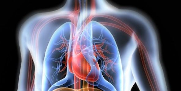
 Cardiopulmonary
Cardiopulmonary
The Cardiopulmonary Department at Atmore Community Hospital is a multifaceted department providing inpatient and outpatient with cardiac and respiratory services. We have Respiratory Therapists here 24 hours a day providing inpatient services to include: oxygen therapy, breathing treatments, arterial blood gas analysis, EKGs, and are also involved in acute and critical care. We also provide inpatient echocardiograms.
Some of the services we provide in our outpatient clinic are EKGs, echocardiograms, pulmonary function testing, 72 hour Holter monitoring and Event monitoring. All tests are required to have a physician's order and can be scheduled by calling our scheduling department.
For more information on specific procedures select the links below:
 Arterial Blood Gas
Echocardiogram (ECHO)
Electrocardiogram (ECG)
Event Monitor
Holter Monitor
Pulmonary Function Test
Arterial Blood Gas
An ABG is a blood test that is performed using blood from an artery. It is a test that measures the pH (acidity/alkalinity), arterial oxygen tension (PaO2), carbon dioxide (PaCO2) and arterial oxyhemoglobin (SaO2). This information is vital when caring for patients with a critical illness or respiratory disease such as COPD or respiratory failure. This test requires an order from the physician but does not need to be scheduled.
Co-Oximetry may be done along with an Arterial Blood Gas (ABG) to help determine the cause of hypoxia or oxygen deficiency at the tissue level. It helps distinguish between carboxyhemoglobin, methemoglobin, oxyhemoglobin and reduced hemoglobin. People who have been exposed to smoke in a house fire or from cigarette smoking can have an increased carboxyhemoglobin which can interfere with the amount of oxygen the patient can have in their blood.
Echocardiogram (ECHO)
An echocardiogram uses sound waves to produce images of your heart which allows the Cardiologist to see how your heart is beating and pumping blood. The Cardiologists can use the images from an echocardiogram to identify various abnormalities in the heart muscle and valves. It gives the Cardiologist information about the size and shape of the heart along with estimates of the heart function such as cardiac output, ejection fraction and diastolic function.
Preparing for the test:
There are few risks involved in a standard echocardiogram. You may feel some discomfort similar to pulling off an adhesive bandage when the technician removes the electrodes placed on your chest during the procedure.
No special preparations are necessary for a standard echocardiogram.
After undressing from the waist up, you'll lie on a bed. The technician will attach electrodes to your chest to help detect and conduct the electrical currents of your heart.
During the echocardiogram, the technician may dim the lights to better view the image on the monitor. You may hear a pulsing "whoosh," which is the machine recording the blood flowing through your heart.
Most echocardiograms take less than 30 minutes, but the timing may vary depending on your condition. During an echocardiogram, you may be asked to breathe in a certain way or to roll onto your left side. Sometimes the transducer must be held very firmly against your chest. This can be uncomfortable - but it helps the technician produce the best images of your heart. A Cardiologist will interpret the echocardiogram and the results will be sent to your doctor.
Electrocardiogram (ECG)
An electrocardiogram is used to monitor your heart. Each beat of your heart is triggered by an electrical impulse normally generated from special cells in the upper right chamber of your heart. An electrocardiogram - also called an ECG or EKG - records these electrical signals as they travel through your heart. Your doctor can use an electrocardiogram to look for patterns among these heartbeats and rhythms to diagnose various heart conditions. An electrocardiogram is a noninvasive, painless test. No special preparations are necessary.
During the test:
Ten electrodes will be attached to your arms, legs and chest. The electrodes are sticky patches applied with a gel to help detect and conduct the electrical currents of your heart. If you have hair on the parts of your body where the electrodes will be placed, the therapist may need to shave the hair so that the electrodes stick properly. You can breathe normally during the electrocardiogram. Moving, talking or shivering may distort the test results. A standard ECG takes just a few minutes.
The ECG is interpreted by a Cardiologist and the results sent to your doctor.
Event Monitor
An event monitor is a heart monitor that is used to record your heart's electrical activity when you have a symptom. You will wear this monitor continuously for up to 30 days. It may be taken off when you shower or bathe. When you feel symptoms you press a button on the monitor and it records the last 45 seconds of and the next 45 seconds of your heart rhythm. The event is then transmitted wirelessly to the monitoring center. When the monitoring center receives your event, if the event shows a rhythm that your physician needs to know about, he/she will be contacted. Depending on the rhythm that is recorded, you may be called and asked to come to the emergency room as soon as possible.
Holter Monitor
A Holter monitor is a small, wearable device that records your heart rhythm. You usually wear a Holter monitor for 72 hours. During that time, the device will record all of your heartbeats. A Holter monitor test is usually performed after a traditional test to check your heart rhythm (electrocardiogram) isn't able to give your doctor enough information about your heart's condition. A Holter monitor has electrodes that are attached to your chest with adhesive and then are connected to a recording device. Your doctor uses information captured on the Holter monitor's recording device to figure out if you have a heart rhythm problem.
Wearing a Holter monitor may be a little inconvenient. Don't swim or bathe for the entire time you're wearing your Holter monitor, although a quick shower is acceptible. While Holter monitors aren't usually affected by other electrical appliances, you shouldn't walk through a metal detector while wearing one. Holter monitoring is painless and noninvasive. You can hide the electrodes and wires under your clothes, and you can wear the recording device on your belt or attached to a strap. Once your monitoring begins, don't take the Holter monitor off - you must wear it at all times, even while you sleep.
Preparing for the test:
A therapist will place an electrode on your chest. For men, a small amount of hair may be shaved to make sure the electrodes stick. If you have hair on the parts of your body where the electrodes will be placed, the therapist may need to shave the hair so that the electrodes stick properly.
The therapist will then connect the electrode to a recording device, so it can record data transmitted from the electrodes.
You'll be instructed to keep a diary of all the activities you do while wearing the monitor. It's particularly important to record in the diary any symptoms of palpitations, skipped heartbeats, shortness of breath, chest pain or lightheadedness.
Once your monitor is fitted and you've received instructions on how to wear it, you can resume your normal activities.
Once your monitoring period is over, you will return to the hospital and a therapist will remove the electrodes from your chest, which may cause some discomfort - similar to a bandage being pulled off your skin. You will turn in the diary you kept while you wore the Holter monitor. The Holter monitor is interpreted by a Cardiologist and the results are sent to your doctor.
Pulmonary Function Test
Pulmonary Function Test (PFT)
A PFT is a diagnostic test that helps the physician identify the severity of lung disease. It can also help determine if the lung disease is reversible by using a medication called a bronchodilator. It measures the ability of the lungs to move large volumes of air quickly in and out of the lungs. All PFT's done are interpreted by a pulmonologist (lung specialist). This test requires an order from the physician and needs to be scheduled prior to being done.